


Brain Imaging Predicts Response to Public Health Campaign
Neuroimaging data obtained from a small group of smokers predicts the influence of a large anti-smoking media campaign targeting likely smokers, shows a new study published in JNeurosci. This approach could help improve informational materials designed to change people's attitudes and behaviors.
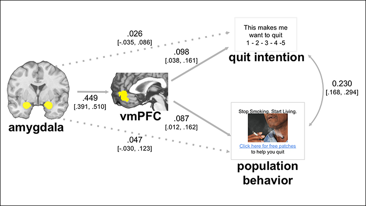
Bruce Dore, Emily Falk and colleagues identified a neural pathway between the amygdala — which is sensitive to emotional content — and the ventromedial prefrontal cortex that predicted the efficacy of graphic anti-smoking messages. The images that smokers said made them want to quit were the same ones that encouraged likely smokers to click through for more information in a New York State Smokers' Quit Line email campaign. Further, images that elicited higher amygdala and vmPFC activity were also more successful in the email campaign — a relationship that was particularly clear when smokers showed low expression of a pattern of brain activity characteristic of emotion regulation. Together these results suggest neuroimaging can be used to predict individual- and population-level responses to persuasive health messages.
Article: Neural mechanisms of emotion regulation moderate the predictive value of affective and value-related brain responses to persuasive messages
Corresponding authors: Bruce Dore, brucedore@gmail.com and Emily Falk, falk@asc.upenn.edu (University of Pennsylvania, Philadelphia, USA)


